
|
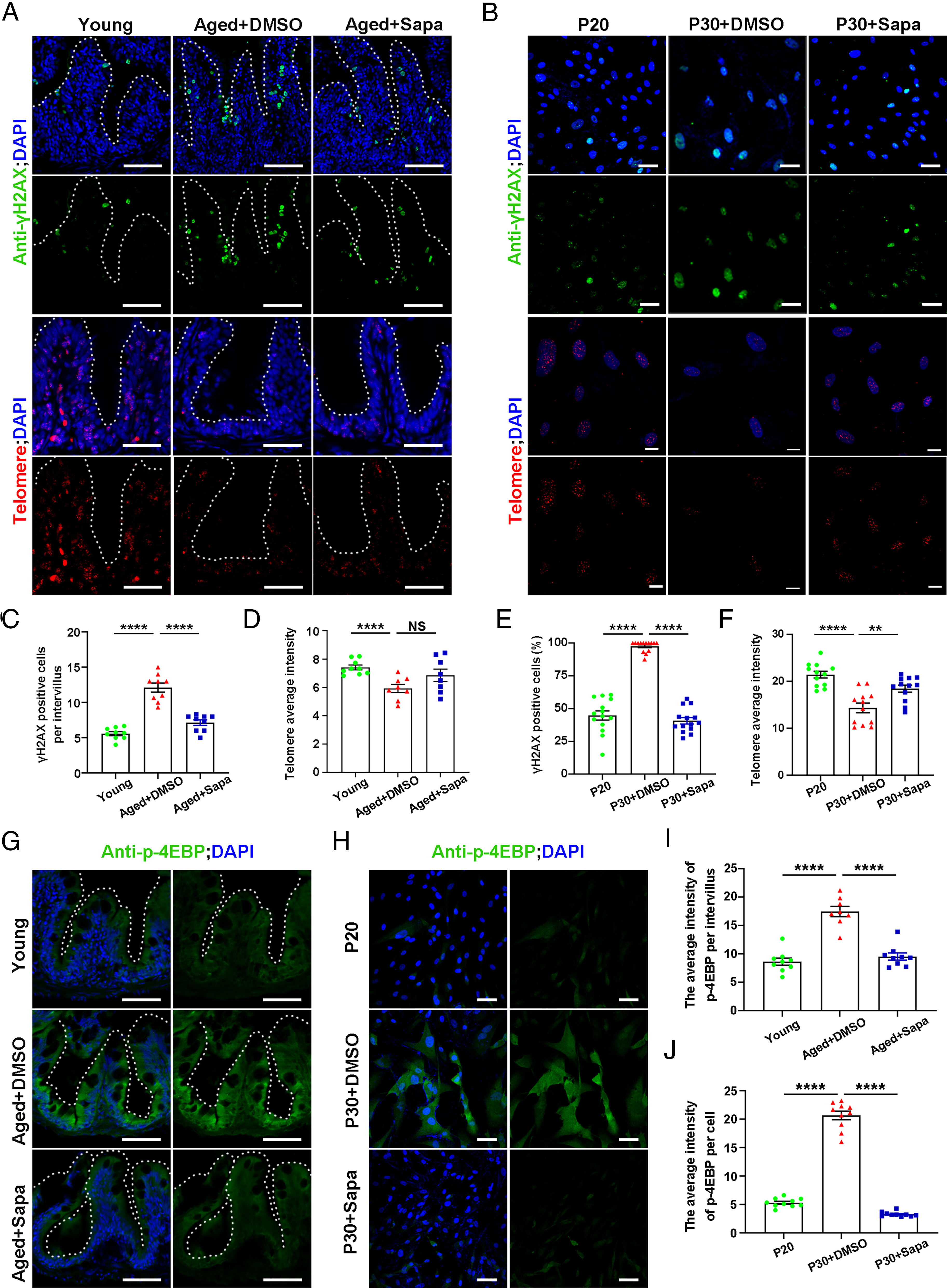
Fig. 6 Sapanisertib attenuated DNA damage and maintained telomeres stability in both physiological-aging zebrafish and replicative-senescent HFF cells via mTORC1 inhibition. (A and B) Representative images of γH2AX staining (Top) and immunofluorescence section images of telomere staining (Bottom) in the intestine of young, aged+DMSO, and aged+Sapa groups (A) and HFF cells of P20, P30+DMSO, and P30+Sapa groups (B). (C–F) Quantification of the γH2AX -positive cells per intervillus in the intestine (C, n = 9 for each group) and in HFF cells (E, n = 14 for each group). Quantification of the intensity of the average fluorescence intensity in the intestine (D, n = 10 for each group) and HFF cells (F, n = 12 for each group). (G–J) Representative images of p-4EBP staining in the intestine (G) and HFF cells (H). Quantification of the intensity of p-4EBP per intervillus in the intestine (I, n = 8 to 9 for each group) and in HFF cells (J, n = 10 for each group). (Scale bar, 100 μm.) Data are presented as mean ± SEM. Significance was determined by one-way ANOVA. NS, not significant; **P < 0.01; ****P < 0.0001. HFF, human foreskin fibroblasts; P20, passage 20.