IMAGE

Figure 5

ID
ZDB-IMAGE-250228-63
Source
Figures for Zheng et al., 2025
Image
Figure Caption

Figure 5

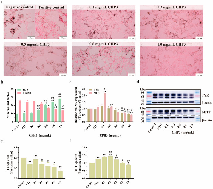
The effect of CPH3 on related protein expression in B16F10 cells under paracrine action. (a) In cultured B16F10, melanin pigmentation was visualized by Fontana–Masson staining. (b) The contents of factors IL-6 and α-MSH in HaCaT cell supernatant were detected by ELISA. (c) Densitometric analysis shows the relative intensity of the nuclear TYR and MITF. (df) Western blotting of TYR and MITF protein level in B16F10. * p < 0.05, ** p < 0.01, versus negative control group. # p < 0.05, ## p < 0.01, versus positive control group (PTU: 1-Phenyl-2-Thiourea, 50 μM), ns: no significant, n = 3.

Acknowledgments
This image is the copyrighted work of the attributed author or publisher, and ZFIN has permission only to display this image to its users. Additional permissions should be obtained from the applicable author or publisher of the image. Full text @ Biomolecules