
Fig. 4
- ID
- ZDB-IMAGE-250227-16
- Publication
- Abello et al., 2024 - Endothelial cell Piezo1 promotes vascular smooth muscle cell differentiation on large arteries
- All Figures
- Figures for Abello et al., 2024
|
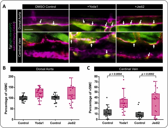
Fig. 4 Activation of Piezo1 activity induces vSMC’s association with the cardinal vein. A) Confocal images of the medial trunk of Tg(tagln:eGFP; kdrl:mCherry-CAAX) zebrafish treated with 10 nM of Yoda1, 400 nM of Jedi2 or DMSO as the vehicle control. vSMCs are shown in green and the endothelium in magenta. Arrows highlight vSMCs associated with the dorsal aorta and cardinal vein. B,C) Quantification of tagln positive vSMCs associated with (B) the dorsal aorta or (C) the cardinal vein following treatment with Yoda1, Jedi2, or DMSO (control). DMSO vs Yoda1 (p = 0.0004; N = 21 and 25); DMSO vs Jedi2 (p = 0.0092; N = 15 and 18). Statistical analysis was performed using a Kruskal-Wallis test with Dunn’s multiple comparison test. Data in B and C were normalized to the control artery condition to allow for collating data from fish across multiple experiments. Scale bar = 50 μm.