
Image
|
Figure Caption
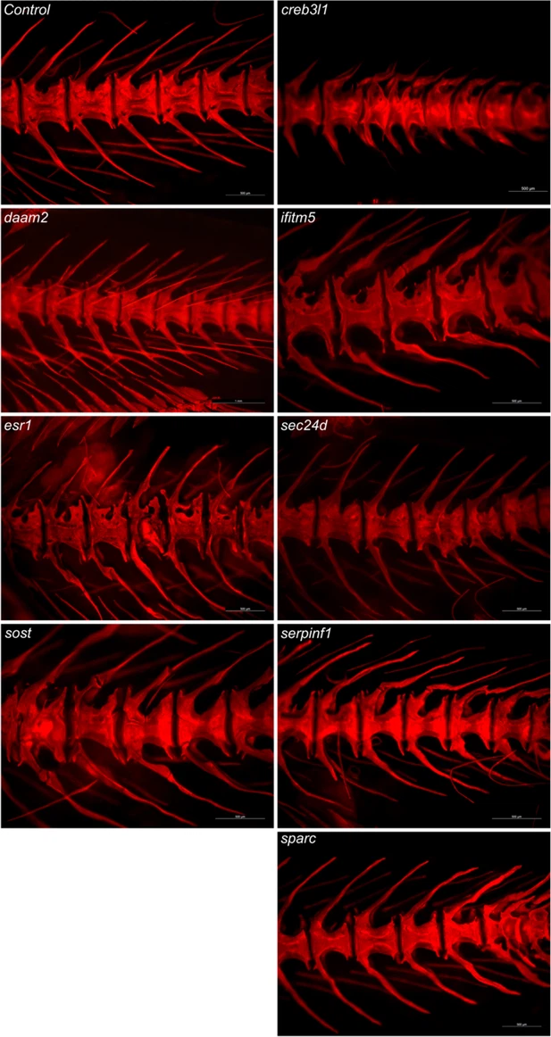
Fig. 4 - Supplemental 3 Mineralization in the skeleton at 90 dpf. Representative images of skeletal mineralization following Alizarin Red S (ARS) staining in a second clutch, demonstrating the consistency of the observed skeletal phenotype. The three genes on the left are associated with the pathogenesis of osteoporosis, while the last five genes on the right are linked to osteogenesis imperfecta. Images show specific crispants from a lateral view, captured using a Leica microscope. Scale bars = 1 mm.
Acknowledgments
This image is the copyrighted work of the attributed author or publisher, and
ZFIN has permission only to display this image to its users.
Additional permissions should be obtained from the applicable author or publisher of the image.
Full text @ Elife