
Image
|
Figure Caption
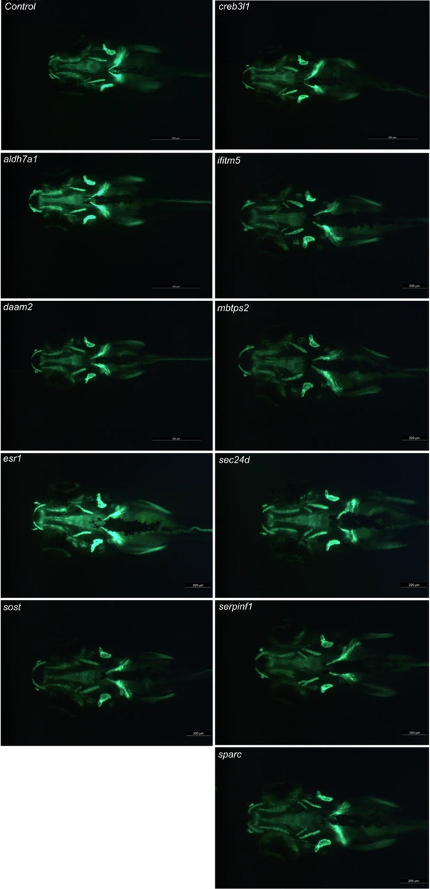
Fig. 1 - Supplemental 2 Osteoblast-positive head area at 7 dpf. Visualization of the osteoblast using the osx:Kaede transgenic line. The four genes on the left are associated with the pathogenesis of osteoporosis, while the six genes on the right are linked to osteogenesis imperfecta. The presented image shows a representative image of the specific crispants. Images are taken with the Leica microscope and the osx:Kaede positive larvae are visualized from a ventral perspective. Scale bars = 500 μm and 200 μm.
Acknowledgments
This image is the copyrighted work of the attributed author or publisher, and
ZFIN has permission only to display this image to its users.
Additional permissions should be obtained from the applicable author or publisher of the image.
Full text @ Elife