IMAGE

Fig. 1

ID
ZDB-IMAGE-250212-58
Source
Figures for Liu et al., 2025
Image
Figure Caption

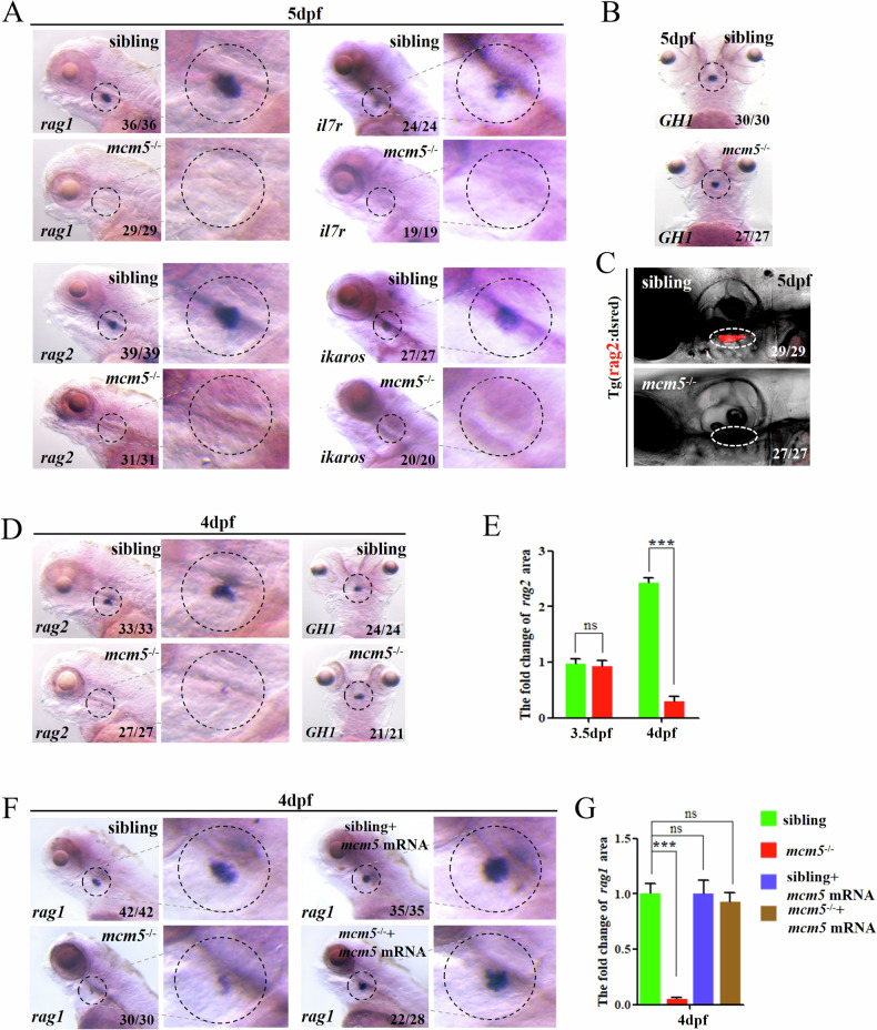
Fig. 1 Mcm5 regulates T lymphocyte maturation during hematopoiesis.

A In control sibling embryos, the expression of rag1, rag2, il7r and ikaros was examined at 5 dpf (shown in the upper-row images). In mcm5−/− embryos, the expression of rag1(n = 29), rag2 (n = 31), il7r (n = 19) and ikaros (n = 20) completely disappeared at 5 dpf (shown in the bottom-row images). BGH1 is a pituitary maker, here working as a control. Comparing to siblings (n = 30), the expression of GH1 in mcm5 mutants was normal (n = 27). C The cells labeled with DsRed (in Tg(rag2:DsRed)) disappeared in mcm5−/− embryos at 5 dpf (n = 27). D Expression of rag2 was greatly decreased in mcm5−/− embryos at 4 dpf (D, n = 27). The expression of GH1 was normal in mcm5−/−embryos at 4 dpf (D, n = 21). E Quantification of the expression area of rag2 in siblings and mcm5−/− embryos at 3.5 dpf and 4 dpf was showed. F The expression of rag1 in mcm5−/− embryos was significantly downregulated at 4 dpf in the thymus (n = 30). Mcm5 mRNA injection restored the expression of rag1 in mcm5−/− embryos (n = 28). G The quantification of the expression area of rag1 at 4 dpf is showed. For (E,G), the data were presented as means ± SD; The P-values (t-test; two-tailed); Ns not significant; *** P < 0.001.

Acknowledgments
This image is the copyrighted work of the attributed author or publisher, and ZFIN has permission only to display this image to its users. Additional permissions should be obtained from the applicable author or publisher of the image. Full text @ Cell Death Dis.