
Image
|
Figure Caption
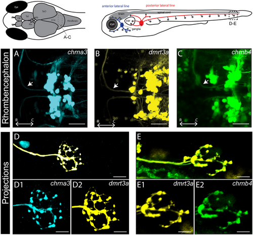
Fig. 3 Expression of chrna3tdTomato, chrnb4eGFP, and chrna5tdTomato in efferent neurons projecting to the lateral line. (A–C) Dorsal view of the hindbrain of chrna3tdTomato, chrnb4eGFP, as well as dmrt3tdTomato expressed in the region where CEN and REN located and the inhibitory efferent axon (arrow) sent out by these cells. (D and E) Projections of chrna3tdTomato and chrnb4eGFP overlapped with dmrt3aeGFP or dmrt3aRFP at the level of neuromasts of the anterior and posterior lateral lines. Scale bars equal 30 µm in (A)–(C), 10 µm in (D) and (E). C, caudal; R, rostral.
Acknowledgments
This image is the copyrighted work of the attributed author or publisher, and
ZFIN has permission only to display this image to its users.
Additional permissions should be obtained from the applicable author or publisher of the image.
Full text @ Dev. Neurobiol.