
|
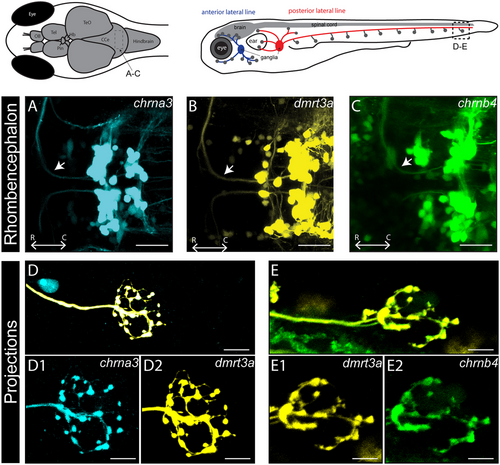
Fig. 2 Expression of chrna3tdTomato, chrnb4eGFP, and chrna5tdTomato in the peripheral nervous system and eyes. (A–C) Lateral view of the intestine of zebrafish larvae at 5 dpf. (A1–C1) Zoom images of marked regions in (A)–(C). (D) Lateral view of the ear labeled by chrnb4eGFP. Dashed circles refer to the outline of ear. (E and F) Lateral view of the eye labeled by chrnb4eGFP and chrna5tdTomato. (G) Lateral view of the eye of transgenic fish of chrna3tdTomato. (H and I) Lateral view of the head labeled by chrnb4eGFP and chrna5tdTomato at 3 dpf revealed the expression in the sensory ganglia, including the trigeminal (gV), facial (gVII), glossopharyngeal (gIX), vagal (gX) ganglia, and both anterior (ALL) and posterior (PLL) ganglia of lateral line. Scale bars equal 60 µm in (A)–(C), 20 in (A1)–(C1), 30 in (D)–(G), 50 µm in (H) and (I). ac, anterior crista; C, caudal; D, dorsal; INL, inner nuclear layer; ipI, inner plexiform layer; Lc, lateral crista; ONL, outer nuclear layer; pc, posterior crista; RGC, retinal ganglion cell.