
|
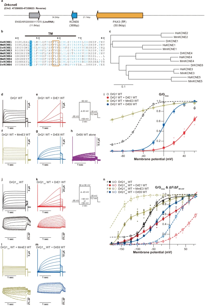
Fig. 1 Biophysical properties of zebrafish KCNE1 and KCNE6 proteins.a Genomic region containing the kcne6 ORF sequence in zebrafish. b Sequence alignment around the TM segments of KCNE1, KCNE3, KCNE4, and KCNE6 proteins. c Phylogenetic tree of KCNE1-6 proteins. Sequence alignment was performed and the phylogenic tree was prepared using the EMBL-EBI Job Dispatcher sequence analysis tools framework63. Sequence alignment is shown using ESPript364. Residues corresponding to “the triplet” 51,52 are highlighted with an orange square. For sequence alignment, human KCNE proteins (HsKCNE1, NCBI Accession Number: NP_000210; HsKCNE2, NP_751951.1; HsKCNE3, NP_005463.1; HsKCNE4, NP_542402.4; and HsKCNE5, NP_036414.1), mouse KCNE proteins (MmKCNE1, NP_001349385.1; MmKCNE2, NP_001345301.1; MmKCNE3, NP_001177798, MmKCNE4, NP_067317.1; and MmKCNE5, NP_067462.1), and zebrafish KCNE proteins (DrKCNE1, XP_017213386.1; DrKCNE4, NP_001076366.1; and DrKCNE6) were used. Representative current traces (d–h) and G-V relationships (i) of DrKCNQ1 WT alone and DrKCNE6 WT alone as well as DrKCNQ1 WT co-expressed with DrKCNE1 WT, MmKCNE3 WT, and DrKCNE6 WT. Representative current traces (upper row) and fluorescence traces (lower row) (j–m) and G-V and F-V (n) of DrKCNQ1vcf WT alone as well as DrKCNQ1vcf WT co-expressed with DrKCNE1 WT, MmKCNE3 WT and DrKCNE6 WT. Error bars indicate ± s.e.m. for n = 5 in (i, n).