
Image
|
Figure Caption
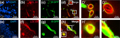
Fig. 7 LDL are taken up by cerebral endothelial cells. (a–h″) intraperitoneal injection of Dil-labelled LDL in Tg(fli1:EGFP) fish (green) allowing to show the vascular distribution of LDLs (in red) in the telencephalon. White squares in d and h identify the high magnifications provided in D′, D″, H′ and H″. The arrows indicate the colocalization of GFP (green) and stained LDL (red), demonstrating that a subset of plasma LDL are taken up by endothelial cells (yellow colour). Bar: 40 μm (a–d), 16 μm (e–h). 10 μm (D′, D″, H′ and H″).
Acknowledgments
This image is the copyrighted work of the attributed author or publisher, and
ZFIN has permission only to display this image to its users.
Additional permissions should be obtained from the applicable author or publisher of the image.
Full text @ Eur. J. Neurosci.