
|
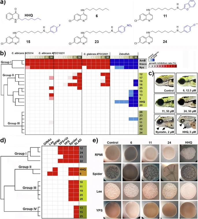
Fig. 1 Identification of the 4-AQ molecules with anti-virulence activity against C. albicans.a The chemical structures of HHQ (2-heptyl-4-hydroxyquinoline) and selected 4-AQ molecules differing in a group bound to the side chain (in blue) and their anti-virulence activities on P. aeruginosa PAO1. b Heatmap illustrating the hierarchical clustering of 4-AQs, HHQ and clinical antifungals, classified using Euclidean distance and Ward’s method into three distinct groups based on their similarity in inhibitory activity against three Candida strains (average of two independent experiments) and toxicity profiles in zebrafish embryos (n = 20 per concentration, tested in triplicate). c The normally developing zebrafish embryos without the signs of toxicity after 5-day exposure to 4-AQ derivatives 11 (Group II), and 6 and 24 (Group III) are shown. In contrast, the embryos treated with 2 µM nystatin (Group I) and 5 µM HHQ were severely damaged and suffered from liver necrosis (dashed line outlining the dark liver), impaired yolk uptake (asterisk), life-threatening pericardial edema (arrow), jaw deformation (arrowhead) and head deformation (bracket). d Heatmap showing the four groups of molecules within the 4-AQs series that differ in their filamentation inhibitory activity (the number of media in which filament formation was completely blocked). After fungal cells were exposed to 10 µM of each molecule for 4-6 days and colony morphology was examined microscopically, each molecule was scored binary for its effect on filament development (presence or absence of filaments) and hierarchically clustered using Ward’s clustering method with Euclidean distance. e The representative images showing the morphology of the fungal colonies after the 3-day treatment with a dose of 10 µM of the selected 4-AQ derivatives (6, 11 and 24). Robust filaments and/or wrinkled colonies were developed in control (0.02% DMSO) treatment, while active molecules inhibited the formation of filaments and/or caused smooth colonies. Hierarchical clustering was performed using the Euclidian distance and Ward’s method.