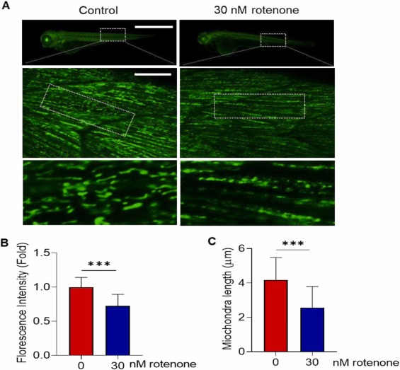
Fig. 7 Rotenone exposure induced mitochondria fragmentation in developing zebrafish embryos. Tg(mito:EGFP) embryos aged 7–8 h post fertilization (hpf) were exposed with 30 nM rotenone to 72 hpf. (A) Representative fluorescence (top) and confocal (middle and bottom) microscopic images. Top scale bar: 1 mm, middle scale bar: 10 µm. Quantification of (B) whole body fluorescence intensity and (C) mitochondria length. Ten mitochondria were randomly selected from the dotted box of the middle image of (A), N = 11–12. Data presented as the mean ± SD. SD: standard deviation. * ** p < 0.001, ns: not significant.
Reprinted from Journal of hazardous materials, 480, Ranasinghe, T., Seo, Y., Park, H.C., Choe, S.K., Cha, S.H., Rotenone exposure causes features of Parkinson`s disease pathology linked with muscle atrophy in developing zebrafish embryo, 136215136215, Copyright (2024) with permission from Elsevier. Full text @ J. Hazard. Mater.

