
Image
|
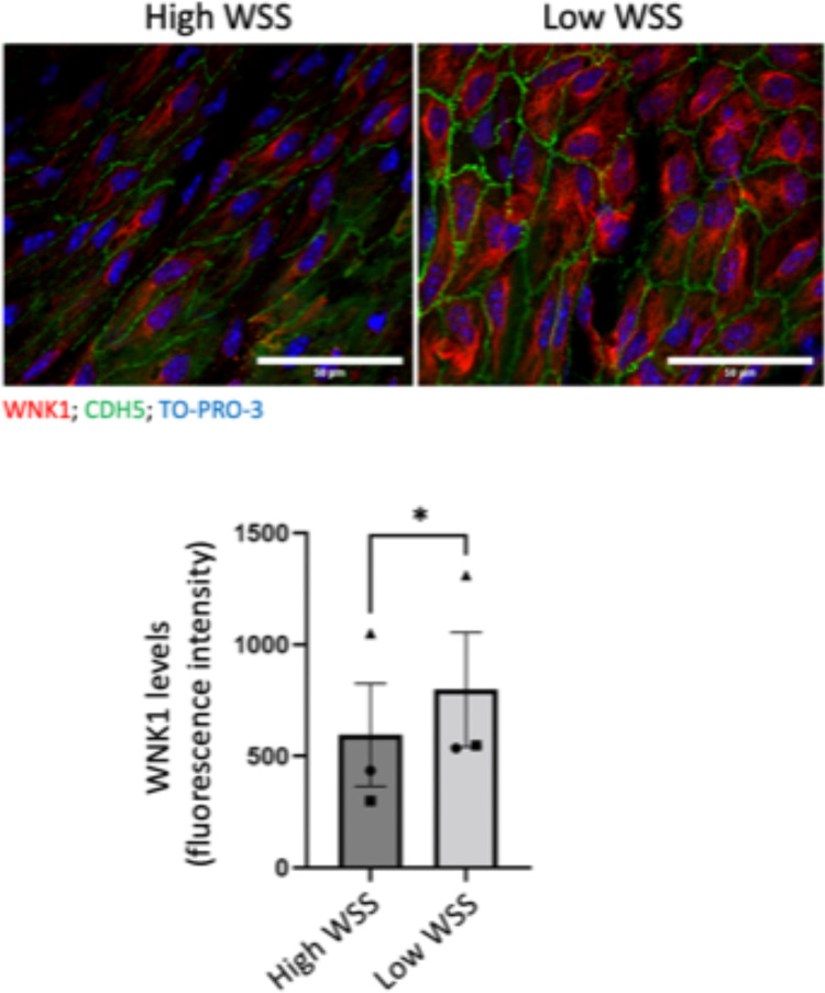
Figure Caption
Fig. 5
WNK1 is enriched at a low WSS region of the mouse endothelium. WNK1 was analysed by en face staining of the murine aorta using anti-WNK1 antibodies and Alexafluor568-conjugated secondary antibodies (red) (n = 3 mice). EC were co-stained using anti-CDH5 (green) and nuclei were detected with TO-PRO-3 (blue). WNK1 expression was quantified in high and low WSS regions. Mean values + /− standard error of mean are shown. Differences between means were analysed using a paired t-test.
Acknowledgments
This image is the copyrighted work of the attributed author or publisher, and
ZFIN has permission only to display this image to its users.
Additional permissions should be obtained from the applicable author or publisher of the image.
Full text @ Sci. Rep.