
Fig. 1.
- ID
- ZDB-IMAGE-241202-31
- Publication
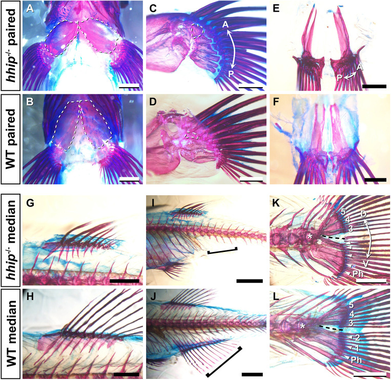
- Tanaka et al., 2024 - Fin elaboration via anterior-posterior constraint by hhip on Hedgehog signaling in teleosts
- All Figures
- Figures for Tanaka et al., 2024
|
Fig. 1.