IMAGE

Fig. 6

ID
ZDB-IMAGE-241123-10
Source
Figures for Li et al., 2024
Image
Figure Caption

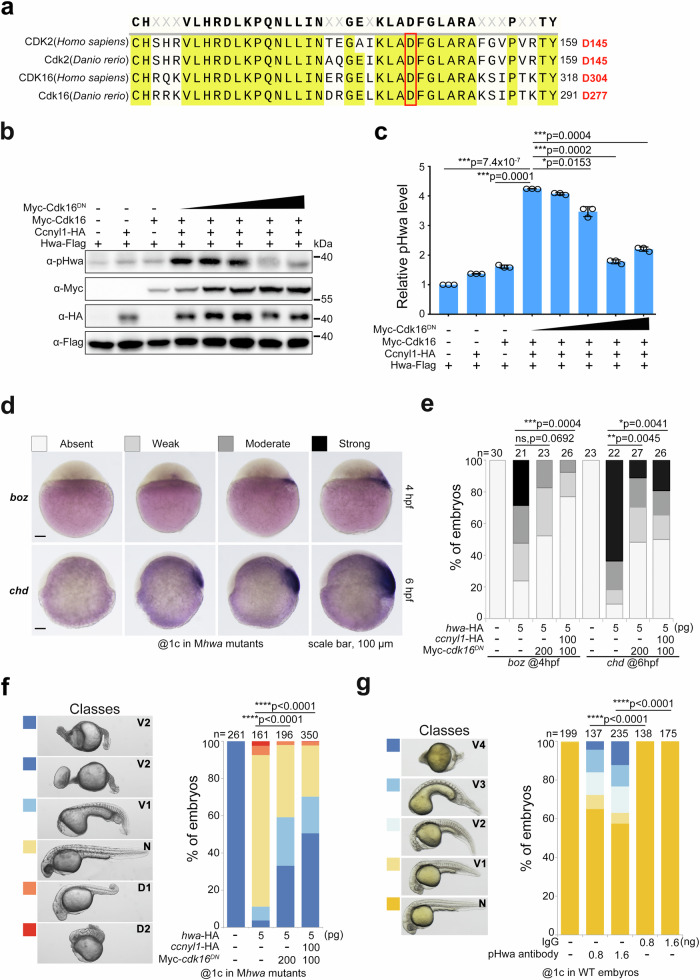
Fig. 6 Attenuating phosphorylation of Ser168 by Cdk16DN or pHwa antibody disrupts the axis-inducing activity of Hwa.

a Residue conservation analysis of human CDK2/16 and zebrafish Cdk2/16 proteins. The red box indicates the conserved, functionally critical aspartic acid (D). b Immunoblotting of pHwa in HEK293T cells transfected with Hwa-Falg, wild-type Cdk16, and different doses of dominant negative Cdk6 (Myc-Cdk16DN). c Quantifications of relative pHwa levels in HEK293T cells treated as in (b), N = 3.Total Hwa protein was used as an internal control. d The WISH results of boz and chd in cdk16DN mRNA injected embryos at 4 hpf and 6 hpf, respectively. e The statistical results of embryos treated as in (d). f Effect of coexpression of cdk16DN and ccnyl1-HA mRNA with hwa mRNA in Mhwatsu01sm embryos, N = 3. g Phenotypes of wild-type embryos injected with pHwa antibody or IgG at different doses, N = 4. Scale bars, 100 μm; V, ventralized; N, normal; D, dorsalized. Total Hwa proteins were used as references for quantification in (c). c A two-tailed unpaired t-test was performed and data were presented as mean ± SD. eg A two-tailed Fisher’s exact test was performed to evaluate differences between treatments (all phenotypes were divided into two groups: Unchanged and Changed). N, number of biological replicates; n, total number of embryos in each treatment; Significant differences are indicated by ns ≥ 0.05, *p < 0.05, **p < 0.01, ***p < 0.001, and ****p < 0.0001. Source data are provided as a Source Data file.

Acknowledgments
This image is the copyrighted work of the attributed author or publisher, and ZFIN has permission only to display this image to its users. Additional permissions should be obtained from the applicable author or publisher of the image. Full text @ Nat. Commun.