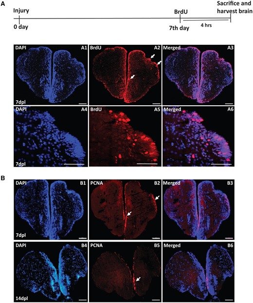
Fig. 4 Immunostaining of regenerating brains showing increased proliferative response upon injury-induced regeneration. (A) Immunostaining of 7 dpl regenerating brain with anti-BrdU (A2) shows increased BrdU-positive cells (white arrows) in the injured/regenerating telencephalic hemisphere. Counterstaining is carried out with DAPI, and A3 shows merged image. (B) Immunostaining of 7 dpl and 14 dpl regenerating brain with anti-PCNA (B2 and B5) shows increased PCNA-positive cells (white arrows) in the injured/regenerating telencephalic hemisphere. Counterstaining is carried out with DAPI. B3 and B6 show the merged images. Scale bar 100 μm.
Image
Figure Caption
Acknowledgments
This image is the copyrighted work of the attributed author or publisher, and
ZFIN has permission only to display this image to its users.
Additional permissions should be obtained from the applicable author or publisher of the image.
Full text @ Biol Methods Protoc

