
|
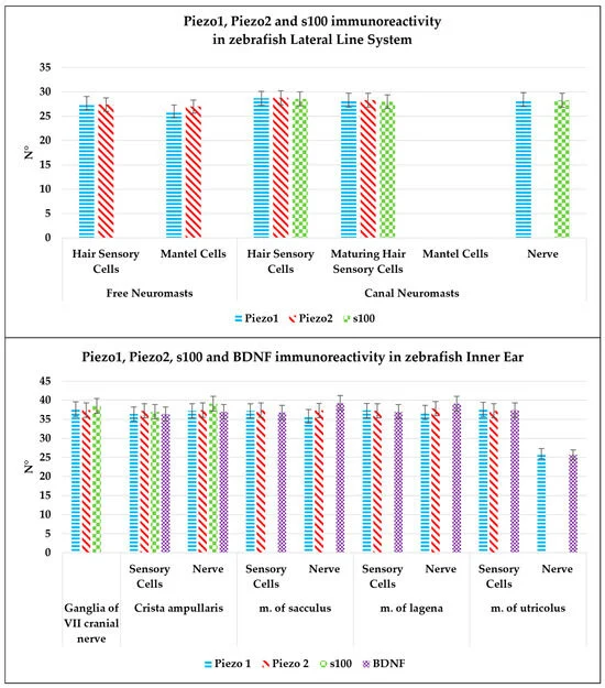
Fig. 8 Graphical representation of immunoreactive cell counts: hair sensory cells and maturing hair sensory cells, mantle cells, and nerve in the neuromast epithelium labeled by Piezo 1, Piezo 2, and s100p; sensory hair cells and nerve of inner ear crista ampullaris and maculae epithelium immunolabeled by Piezo 1, Piezo 2, BDNF, and s100p; neurons of VIII cranial nerve immunostained with Piezo 1, Piezo 2, and s100p. The statistical analysis shows a different expression pattern of the investigated protein in different cellular subpopulations. N°: mean of cells immunopositive to Piezo 1, Piezo 2, BDNF, and s100p. The significant difference was assessed for p < 0.05.