
|
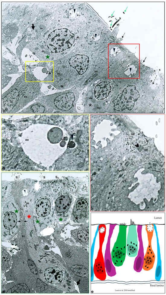
Fig. 1 Transmission electron micrograph of a transverse section of an adult zebrafish (D. rerio) canal neuromast. Heterogeneity among sensory hair cells (HCs) can be observed. (a) A HC with a light cytoplasm (green star) and HCs with a more dense, strongly stained cytoplasm (asterisks), both with sparce heterochromatin nucleus (n), vesicle (v), and numerous electron-dense mitochondria (m), are evident. Note an HC showing a group of stereocilia with a typical staircase arrangement (st) in addition to a detached kinocilium (K) and its cross-sections (green arrows). Junction complexes between an HC and a support cell (sc) are indicated by a red inset. At the basal pole of the sensory hair cells, afferent (yellow inset) and efferent (thick arrow) synapses are visible. The occurrence of maturing HCs close to the apical surface is characterized by a peculiar crypt-like rounded space (arrowheads), with stereocilia (s). A maturing volcano-like HC, already reaching the neuromast lumen, with a peculiar depression (broken-arrow) and with the stereocilia surface projecting from the cell surface (s) to the lumen is identified. Elongated scs underneath or close to the HC, sending thin cytoplasmic projections apically, are evident. Note some microvilli (thin arrows) on the top of the sc. (b) Higher magnification of the basal pole of a sensory hair cell. Afferent synapse (aa) characterized by a classical pre-synaptic body (arrowhead) and a post-synaptic side with a clear cytoplasm and mitochondria (m) are visible. (c) Higher magnification of the apical surface of an HC and an sc. Zonula occludens at the apical surface (broken arrow) and more basally desmosomal-like junctions between the HC and sc are clearly visible (gallon arrow). Mitochondria (m). (d) Numerous HCs with dense (asterisk) and lighter cytoplasm (green stars) placed close to the neuromast apical surface. The occurrence of stereocilia (s) in the apical part of the HC. The occurrence of a maturing pear-shaped HC (red star) extending to the apical surface from the basal lamina to the apical surface of the neuromast. Note at the apical pole a peculiar, rounded space with some stereocilia inside (arrowhead). Note the distribution of heterochromatin in the nucleus (n). Mitochondria (m). Afferent synapse (arrow). sc with cytoplasmic projections (in between arrows) separating two adjacent HCs and extending to the apical surface. (a,d) 5000×, (b,c) 10,000×. (e) A graphical representation of the cell subpopulations of the neuromast sensory epithelium: mantle cells (blue), maturing crypt-like HC (red cell), sc (purple cells), hair cells with dense cytoplasm (green cell), hair cells with light cytoplasm (light green cell), maturing volcano-like HC (orange) (Laurà et al. [29] modified).