
Fig. 5
- ID
- ZDB-IMAGE-240916-121
- Publication
- Williams et al., 2024 - Keratin 8/18a.1 Expression Influences Embryonic Neural Crest Cell Dynamics and Contributes to Postnatal Corneal Regeneration in Zebrafish
- All Figures
- Figures for Williams et al., 2024
|
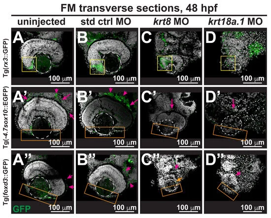
Fig. 5 Krt8/Krt18a.1 play roles in neural crest migration and patterning during early eye development. Section analysis of 48-hpf Tg(-4.7sox10::EGFP), Tg(foxd3::GFP), and Tg(rx3::GFP) zebrafish embryos showed that MO knockdown of K8 or K18a.1 resulted in the significant disorganization of neural crest cells in the optic cup rim (yellow box, (C,D)), periocular mesenchyme (magenta arrow, (C′,D′,C″,D″)), and ocular anterior segment (orange box, (C′,D′,C″,D″)). The orange arrow indicates slight foxd3 signal in the anterior segment of the underdeveloped eye. The dashed circles highlight the lens. (E) Quantitative analysis showed a significant decrease of rx3, sox10, and foxd3 promoter-driven GFP expression in the periocular and ocular regions of the developing eyes of krt8/krt18a.1 morphants compared with those of uninjected (A–A″) and control MO-injected fish (B–B″). Notably, these effects were more severe with K18a.1 knockdown. **, p-value < 0.01; n.s., not significant.