
|
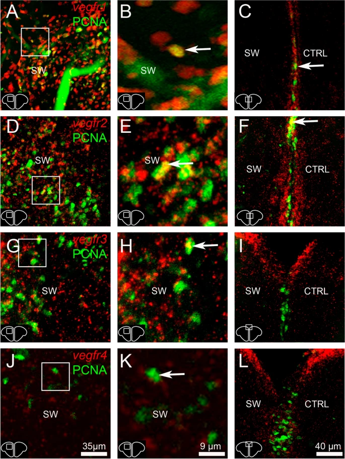
Fig. 14 Numerous proliferative cells in the parenchyma are vegfr-positive. A, B, D, E, G, H, J and K vegfr1, vegfr2, vegfr3, and vegfr4 (red) in situ hybridization followed by PCNA immunohistochemistry (green) at 3 dpl in the stabwounded (SW) hemisphere. Arrows show examples of proliferative cells expressing vegfr. C, F, I and L vegfr1, vegfr2, vegfr3, and vegfr4 (red) in situ hybridization followed by PCNA immunohistochemistry (green) at 5 dpl in the ventricular zone containing neural stem cells that actively proliferate following injury at this time. Most of proliferative cells did not express vegfr (or barely), except for some of them (see arrows). Scale bar = 35 µm (A, D, G and J), 9 µm (B, E, H and K), 40 µm (C, F, I and L)