
|
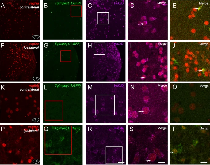
Fig. 5 vegfaa and vegfbb are up-regulated after brain lesion notably in neurons and microglia. Fluorescent ISH (red) followed by HuC/D immunostaining (purple) on Tg(mpeg1.1:GFP) (green) showing vegfaa and vegfbb at 1 day post-lesion in contralateral and ipsilateral telencephalic hemispheres. (D, I and E, J) High magnification views at the levels of the white and red squares showing vegfaa expression in neurons (D and I) and in microglia (E and J). (N, S and O, T) High magnification views at the levels of the white and red squares showing vegfbb expression in neurons (N and S) and in microglia/immune cells (O and T). Bar: 50 µm (A-C and F–H), 14 µm (D-E, I-J, K-M and P-R), 7 µm (N–O and S-T)