
Image
|
Figure Caption
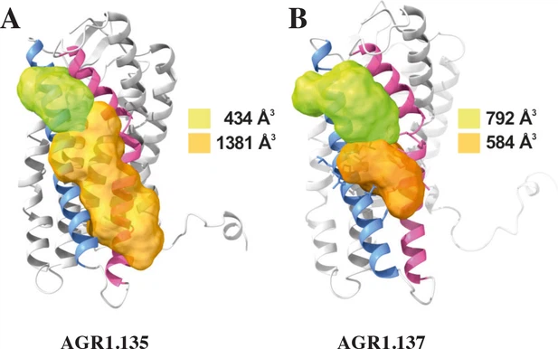
Fig. 6 - supplement 1 Cartoon and surface representation of CXCR4 and the clefts identified between TMV and TMVI. The protein structure is shown in gray, with TMV and TMVI colored in blue and pink, respectively. Cavities associated to AGR1.135 (A) and AGR1.137 (B) binding were identified by SurfNet software are shown in orange and green. Volumes for each cavity are measured (Å3).
Acknowledgments
This image is the copyrighted work of the attributed author or publisher, and
ZFIN has permission only to display this image to its users.
Additional permissions should be obtained from the applicable author or publisher of the image.
Full text @ Elife