
|
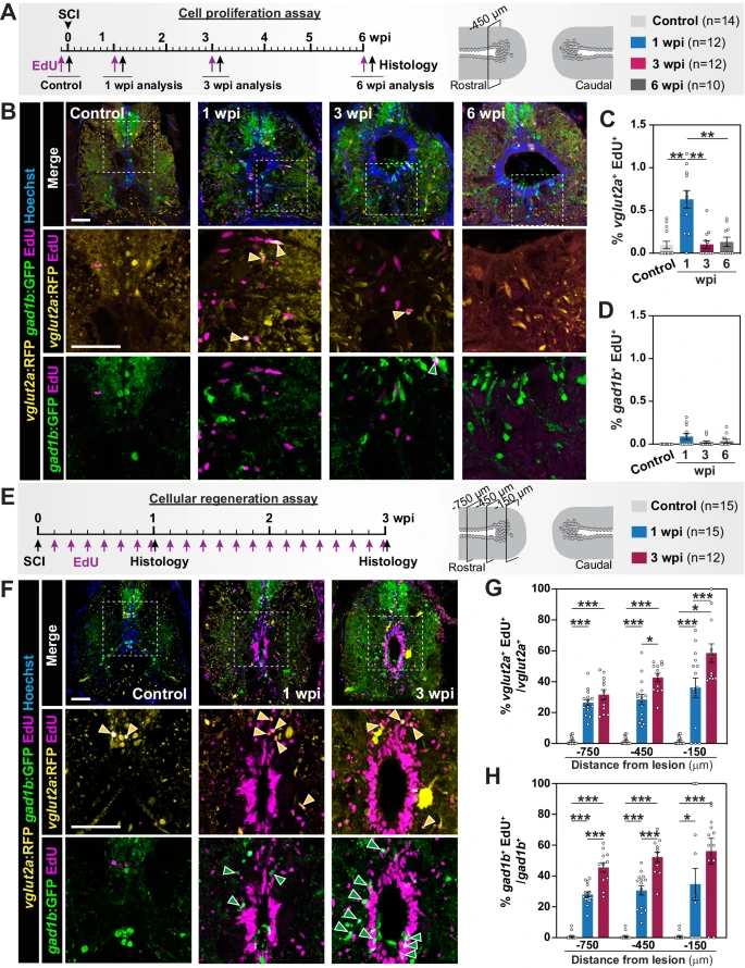
Fig. 4 Regeneration of excitatory and inhibitory neurons after spinal cord injury. A Experimental timeline to assess cell proliferation. SC cross-sections 450 µm rostral to the lesion were used for quantification. B Staining for RFP, GFP, EdU and Hoechst in Tg(vglut2a:RFP; gad1b:GFP) zebrafish at 0, 1, 3 and 6 wpi. Insets marked by dotted lines represent dual channels for RFP and EdU or for GFP and EdU. Yellow arrowheads indicate vglut2a+ EdU+ neurons. Green arrowheads indicate gad1b+ EdU+ neurons. C, D Quantification of vglut2a+ EdU+ neurons (ANOVA p < 0.0001) and gad1b+ EdU+ neurons (ANOVA p: 0.0449) at 0, 1, 3 and 6 wpi. Percent cells were normalized to the total number of nuclei in SC sections. E Experimental timeline to assess the cumulative profiles of regenerating neurons at 1 and 3 wpi. Tissue sections 150, 450 and 750 μm rostral to the lesion were analyzed. F Staining for RFP, GFP, EdU and Hoechst in Tg(vglut2a:RFP; gad1b:GFP) zebrafish at 0, 1 and 3 wpi. Insets marked by dotted lines represent dual channels for RFP and EdU or for GFP and EdU. Yellow arrowheads indicate vglut2a+ EdU+ neurons. Green arrowheads indicate gad1b+ EdU+ neurons. G, H Quantification of regenerating glutamatergic neurons (vglut2a+ EdU+) (ANOVA p < 0.0001) and regenerating GABAergic neurons (gad1b+ EdU+) (ANOVA p < 0.0001). For each section, percent cells were normalized to the number of vglut2a+ neurons in (G) and to gad1b+ neurons in (H). Data points in all bar charts indicate individual animals and sample sizes are indicated in parentheses. Brown-Forsythe and Welch ANOVA were performed in (C, D) with Dunnett’s T3 multiple comparisons performed across time points with 95% CI. Two-way ANOVA was performed in (G, H) with Tukey’s multiple comparisons with 95% CI. In all bar charts, data are presented as mean values +/- SEM. All statistical tests are two-sided. *p ≤ 0.05, **p ≤ 0.01, ***p ≤ 0.001. Scale bars, 50 µm. Source data are provided as a Source Data file.