
|
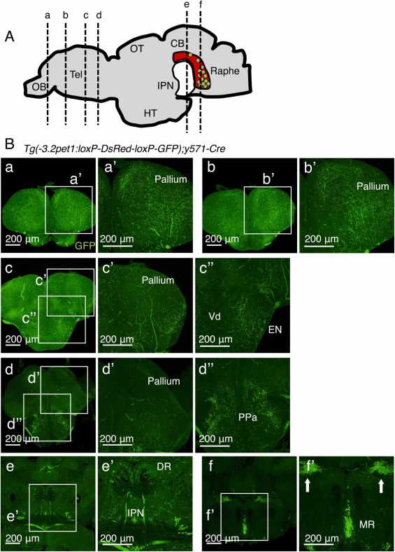
Fig. 3 The median raphe serotonergic neurons derived from rhombomere 2 projected axons to the rostral dorsal pallium. (A) Schematic diagram of the position of the coronal sections. (B) Panel a, and a’ are the most anterior section of the telencephalon. Panels b, and b’ are sections of the anterior telencephalon. Panels c and c’ are sections of the posterior telencephalon. Panels d and d’ are the most posterior telencephalon sections. Panels e and e’ are sections of the dorsal raphe. Panels f and f’ are sections of the median raphe. The white arrows in the panel f’ indicate the axonal signals of GFP. The age of zebrafish in this experiment is 15 months.