
|
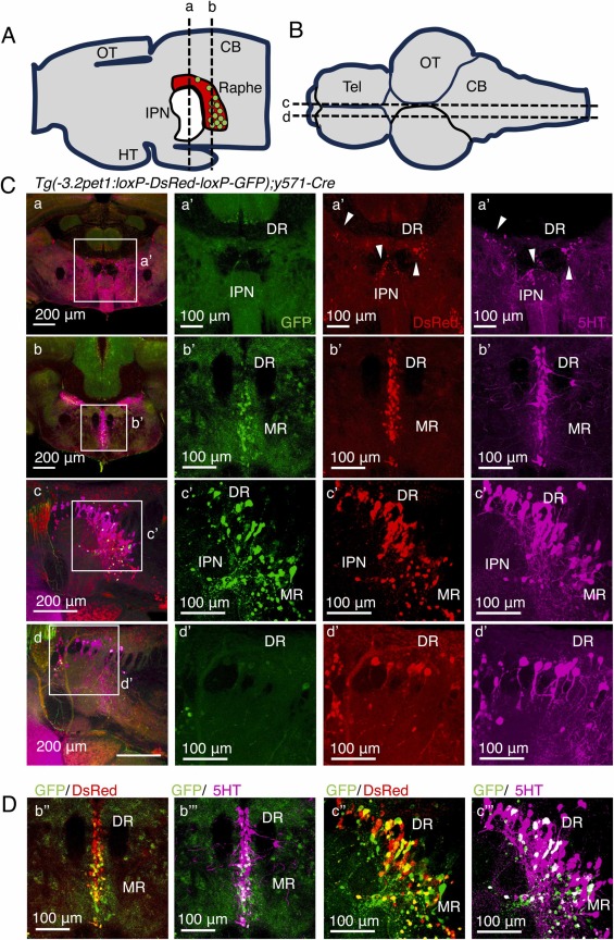
Fig. 2 Localization by immunohistochemistry of GFP and DsRed in Tg(-3.2pet1:loxP-DsRed-loxP-GFP);y571-cre. (A, B) The positions of a cross-sections for immunohistochemistry (A, coronal; B, sagittal) (C) Fluorescence results of immunohistochemistry of GFP (Green), DsRed (red), and 5HT (magenta). Panels a, and a’ are coronal sections, including the IPN and the dorsal raphe. Panels b and b’ are coronal sections, including the dorsal and median raphe. Panels c and c’ are sagittal sections including the IPN, the dorsal raphe, and the median raphe. Panels d, and d’ are sagittal sections, including the lateral side of the dorsal raphe. (D) Panels b’’ and c’’ are merged images with GFP (green) and DsRed (red). Panels b’’’ and c’’’ are merged images with GFP (green) and 5HT (magenta). The age of zebrafish in this experiment is 4 months.