
|
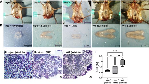
Fig. 3 Gonadal morphology and gametogenesis of vipa−/− and WT testes. Gross testes morphology (in situ) of 2 representative vipa−/− and WT adult males (A; top row), and after excision (B; bottom row). (C) Testis histology with hematoxylin and eosin staining of vehicle-treated vipa−/− male. (D) Testis histology with hematoxylin and eosin staining of methyl-testosterone treated vipa−/− male. (E) Testis histology with hematoxylin and eosin staining of WT male. (F) Average GSI of WT and vipa−/− males (n = 6). Statistical analysis performed using the Welch 2-sample t-test. P (GSI) = .0092 and .0002, **P < .01, ***P < .001.