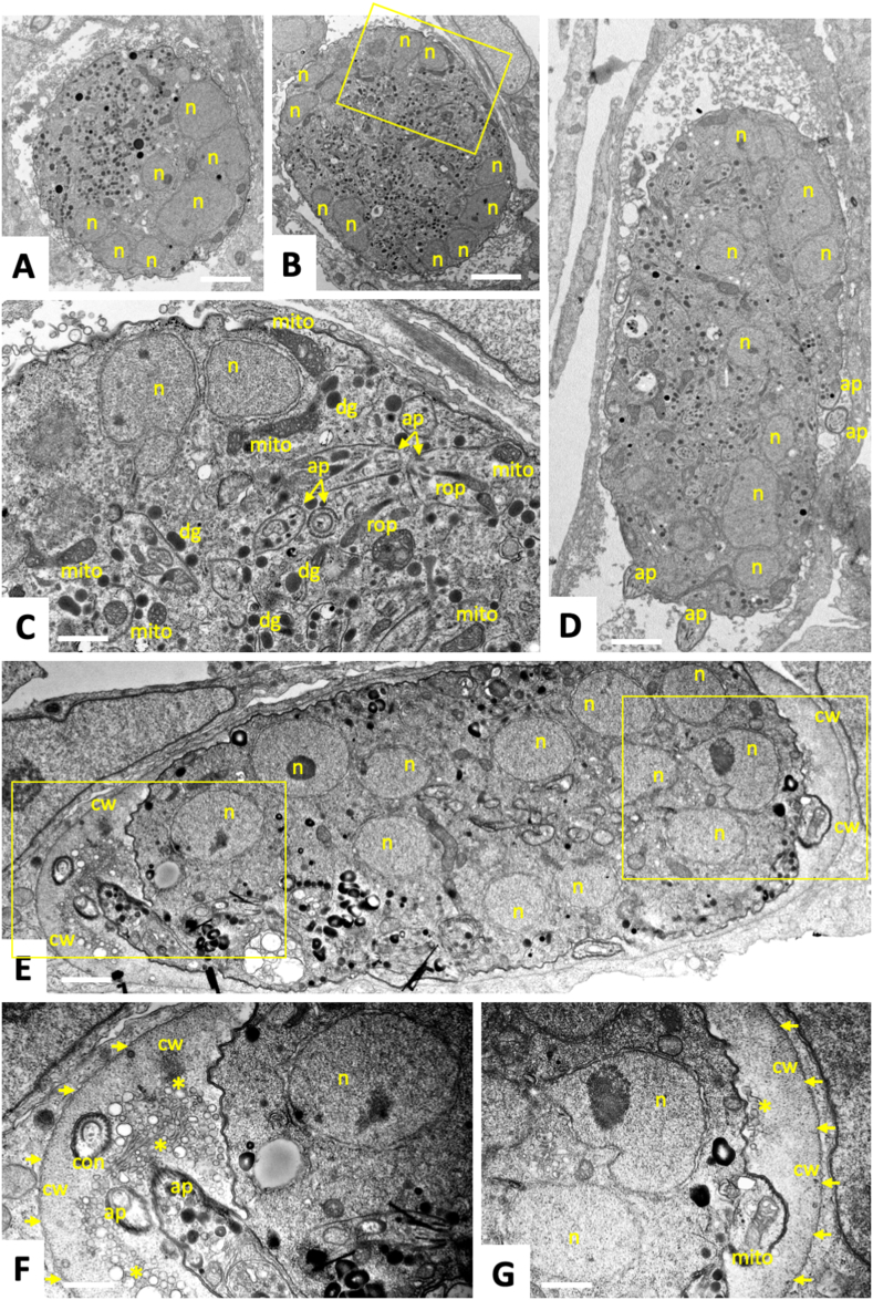
IMAGE

Fig. 3

ID
ZDB-IMAGE-240720-4
Source
Figures for de Sousa et al., 2024
Image
Figure Caption

Fig. 3

Transmission Electron Microscopy of T. gondii tachyzoites grown in human foreskin fibroblasts and treated with BKI-1708 (2.5 μM) during 48 h (A–C), 72 h (D) and 96 h (E–G). The boxed area in (B) is enlarged in (D), the two in (E) are enlarged in (F) and (G); n = nucleus, ap = apical part of newly formed zoite, dg = dense granules, rop = rhoptries, mito = mitochondrion, con = conoid, cw = cyst wall like structure. Small arrows point towards the parasitophorous vacuole membrane, * indicate secretory vesicles. Bars in (A) and (B) = 2.1 μm; (C) = 0.6 μm; (D) = 2.4 μm; (E) = 1.2 μm; (F) = 0.7 μm and (G) = 0.5 μm.

Acknowledgments
This image is the copyrighted work of the attributed author or publisher, and ZFIN has permission only to display this image to its users. Additional permissions should be obtained from the applicable author or publisher of the image. Full text @ Int J Parasitol Drugs Drug Resist