
Fig. 2
- ID
- ZDB-IMAGE-240716-14
- Antibodies
- Publication
- Lu et al., 2024 - Localisation and function of key axonemal microtubule inner proteins and dynein docking complex members reveal extensive diversity among vertebrate motile cilia
- All Figures
- Figures for Lu et al., 2024
|
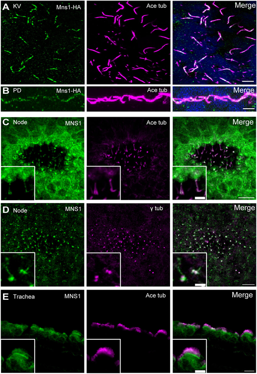
Fig. 2 MNS1 localisation to motile cilia in zebrafish and mouse tissues. (A) Immunofluorescence with antibodies to HA epitope (green) and acetylated tubulin (magenta) of cilia in zebrafish KV [10-somite stage (14 hpf)]. (B) Immunofluorescence with antibodies to HA (green) and acetylated tubulin (magenta) of zebrafish pronephric duct (PD) cilia (24 hpf). (C) Immunofluorescence with antibodies to MNS1 (green) and acetylated tubulin (magenta) of E8.0 mouse node cilia. (D) Immunofluorescence with antibodies to MNS1 (green) and γ tubulin (magenta) of E8.0 mouse node cilia. (E) Immunofluorescence with antibodies to MNS1 (green) and acetylated tubulin (magenta) of adult mouse trachea. Insets in C-E show higher magnification views. Scale bars: 5 µm (A,B,E, main panel); 2 µm (C,D, main panels); 10 µm (C-E, insets).