
Fig. 6
- ID
- ZDB-IMAGE-240715-6
- Publication
- Maeno et al., 2024 - Hox code responsible for the pattering of the anterior vertebrae in zebrafish
- All Figures
- Figures for Maeno et al., 2024
|
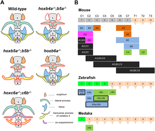
Fig. 6 Hox genes responsible for the anterior vertebral identities in mouse, zebrafish and medaka. (A) Diagrams illustrating the typical morphologies of the anterior vertebrae in the zebrafish Hox mutants isolated in this study. The bones that are observed to be abnormal in Hox mutants are highlighted in red, and the missing bones are indicated by dotted lines. Ventral view. (B) Comparisons of Hox genes responsible for the anterior vertebrae in mouse, zebrafish and medaka. Each vertebra is represented by a box, and a triangle attached to the box indicates thoracic vertebrae with ribs. In mice, Hox genes that define the vertebral identities are shown based on malformed vertebrae in Hox knockout mice. In mice, Hox genes from paralog 3 are responsible for vertebral patterning. The specific Hox gene is shown below the vertebrae that are abnormal in individual Hox knockout mice: Hoxb3 (Manley and Capecchi, 1997), Hoxd3 (Condie and Capecchi, 1993), Hoxa4 (Horan et al., 1994; Kostic and Capecchi, 1994), Hoxb4 (Ramirez-Solis et al., 1993), Hoxc4 (Saegusa et al., 1996), Hoxd4 (Horan et al., 1995a), Hoxa5 (Jeannotte et al., 1993), Hoxb5 (Rancourt et al., 1995), Hoxa6 (Kostic and Capecchi, 1994), Hoxb6 (Rancourt et al., 1995) and Hoxc6 (Garcia-Gasca and Spyropoulos, 2000). Multiple paralogous Hox knockout mice showed more severe vertebral abnormalities than single knockout mice. For paralog 3, which has three paralogs, no triple Hox mutants have been reported, but double mutants combining two of the three mutations showed vertebrae with abnormalities (Condie and Capecchi, 1994; Manley and Capecchi, 1997). For paralog 4, triple mutants were reported among the four paralogs, showing C2-C5 vertebrae with abnormalities (Horan et al., 1995b). For paralogs 5 and 6, triple mutants lacking all paralogs have been reported with abnormal vertebrae (McIntyre et al., 2007). In zebrafish, Hox genes that define specific vertebrae, as revealed in this study, are shown below the vertebrae. The frame of the box is black where vertebral abnormalities were observed in double Hox mutants. In medaka, hoxc6a is responsible for the second vertebra.