
|
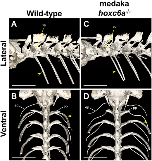
Fig. 5 The second vertebra of medaka hoxc6a−/− mutants shows abnormalities without ribs, similar to the first vertebra. (A-D) Micro-CT scan analysis of the anterior vertebrae in wild-type (n=3) and hoxc6a−/− (n=3) adult medaka. As similar phenotypes were observed for all mutants of the same genotype, a representative individual is shown. The arrowheads indicate the anterior-most pleural ribs attached to vertebrae. In wild-type medaka, the ribs are present from the second vertebra (n=3), but in hoxc6a mutants, the ribs are observed from the third vertebra (n=3). Micro-CT scan 3D movies are provided in Movies 14,15. Scale bars: 1 mm. ep, epipleurals; pp, parapophysis.