
|
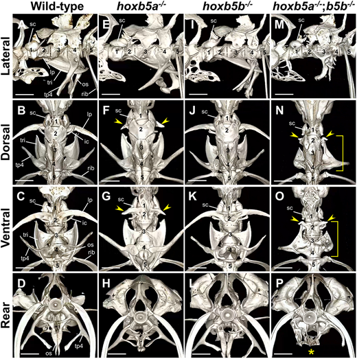
Fig. 3 Severe defects in the second to fourth vertebrae of zebrafish hoxb5a−/−;hoxb5b−/−mutants. (A-P) The anterior vertebrae were examined using micro-CT scan in the following groups: wild-type (A-D, n=4), hoxb5a−/− (E-H, n=4), hoxb5b−/− (I-L, n=3) and hoxb5a−/−;hoxb5b−/− (M-P, n=4) adult fish. Representative images are shown. Fish of the desired genotype were obtained by several intercrosses between hoxb5a+/−;hoxb5b+/− fish and the genotype analysis of the surviving juveniles is shown in Table S1C. The numerical values correspond to the positional order of each vertebra from the first vertebra. The arrowheads indicate a significantly shortened lateral process on the second centrum in hoxb5a−/− and hoxb5a−/−;hoxb5b−/− mutants. The brackets indicate malformation of the ossicles on the second to fourth centrum in hoxb5a−/−;hoxb5b−/− mutants. The asterisk indicates the absence of the os suspensorium in hoxb5a−/−;hoxb5b−/− mutants. Scale bars: 1 mm. For additional details, micro-CT scan 3D movies are provided in Movies 8,9,10. A summary of the phenotypes of these mutants is shown in Table S2. Digital dissection of each vertebra in hoxb5a−/−;hoxb5b−/− is shown in Fig. S3. ic, intercalarium; lp, lateral process; os, os suspensorium; sc, scaphium; tp4, transverse process of vertebra 4; tri, tripus