
|
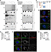
Fig. 3 ARMC9 interacts with NDUFAF2 via its C-terminal region, and their interaction is required for localization of mitochondria and centrosome. (A and B) 293T cells transfected with various expression constructs analyzed by immunoprecipitation followed by Western blots with indicated antibodies. (C) Analysis of 293T cells transfected with various expression constructs by immunoprecipitation followed by Western blots. Schematic diagram showing various ARMC9 mutants tagged with FLAG. The ability of each construct to interact with NDUFAF2 is also indicated. CC, mean coiled-coil domain; LisH, mean predicted lissencephaly type 1–like homology motif. (D) Cells were incubated with a centriole marker (centrin, green) and with anti-ARMC9 and anti-NDUFAF2 antibodies and then probed using PLA Minus anti-ARMC9 and Plus anti-NDUFAF2 (red). Scale bars: 10 μm, 2 μm. (E) Quantification of puncta around centrioles in wild-type, NDUFAF2–/–, and NDUFAF2WT-re-expressing RPE1 cells. (F) Cells were incubated with a mitochondrial marker (green) and with anti-ARMC9 and anti-NDUFAF2 antibodies and then probed using PLA (red). Scale bar: 10 μm. (G) Quantification of puncta on mitochondria in wild-type, NDUFAF2–/–, and NDUFAF2WT-re-expressing RPE1 cells. The bars in each graph represent mean ± SD. Exact P values are indicated. ANOVA followed by Tukey-Kramer multiple-comparison test.