
Image
|
Figure Caption
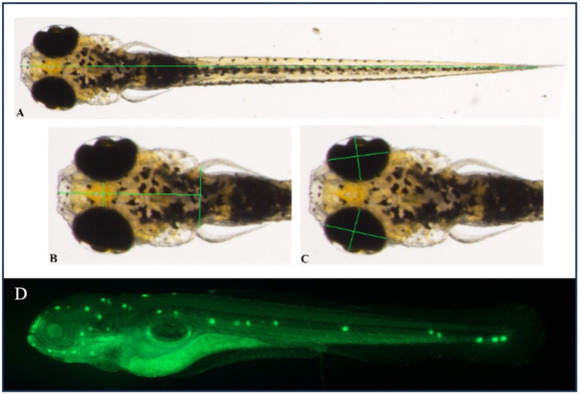
Fig. 2 Illustrative photographs (dorsal view) and markings of the morphological parameters analyzed for body, cranial, and ocular measurements of zebrafish larvae at the 5 dpf stage: body length (1A) at 4 × magnification; length of head and interocular distance (1B) at 6 × magnification; ocular height and width (1C) at 6 × magnification. D) Image showing the apoptotic cells using the acridine orange technique. Islands of apoptotic cells can be seen with fluorescent labeling. Photographs were taken with a stereomicroscope; side view, 4 × magnification.
Acknowledgments
This image is the copyrighted work of the attributed author or publisher, and
ZFIN has permission only to display this image to its users.
Additional permissions should be obtained from the applicable author or publisher of the image.
Full text @ Heliyon