
|
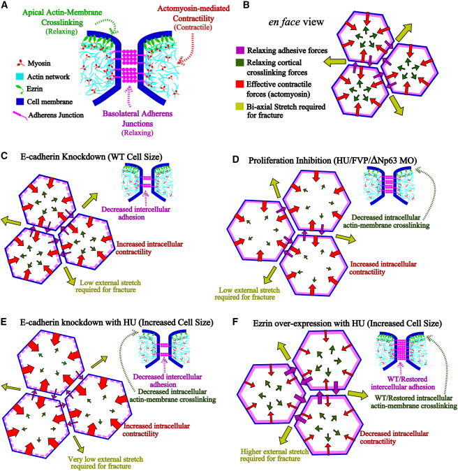
Fig. 6 Ezrin and E-cadherin counter myosin-II activity to maintain effective contractility and epidermal resilience under tensile stress (A) Schematic depicts the experimentally determined localization of force generating complexes in the cell. The actin-membrane crosslinking via Ezrin is present at the apical cortex, while adherens junctions and the associated actomyosin pool localize to the basolateral domain. (B) Schematic depicts various forces acting on the wild-type epithelial cells that balance each other and determine the biaxial stretch necessary to fracture the tissue (yellow arrows). Here, the actomyosin-mediated effective contractile forces are depicted in red arrows, while the counteracting forces due to Ezrin and E-cadherin-mediated relaxation are depicted in green and purple arrows, respectively. (C–F) Schematics show how different perturbations to either cell adhesion (C), cell size (D), or both (E) affect this force balance and decrease the maximum stretch tolerated by the epithelium prior to fracture. The schematic in (F) shows the importance of reinforcement of both cell-cell adhesion and cortical crosslinking in restoring effective tissue contractility and increasing fracture strength under proliferation inhibition. Note that the thickness of the arrows represents their scalar magnitudes.