
|
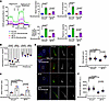
Fig. 5 NAD+ supplementation rescues defective ciliogenesis in JBTS patient-derived cells. (A) OCR measured by Seahorse Analyzer of HConF cells and JBTS cells with or without nicotinamide treatment. (B) Analysis of mitochondrial complex I activity in HConF cells, JBTS cells, and both cell lines overexpressing NDUFAF2. (C) Analysis of the NAD+/NADH ratio in HConF cells and JBTS cells. (D) Immunostaining of cells serum-starved for 2 days. Scale bars: 10 μm, 1 μm. (E) Quantification of cilia length in HConF cells and JBTS cells treated with nicotinamide; >50 cells analyzed for each independent experiment. (F) Quantification of NPHP1 signal intensity at the centrioles; >100 cells analyzed for each independent experiment. The bars in each graph represent mean ± SD. Exact P values are indicated. ANOVA followed by Tukey-Kramer multiple-comparison test.