
|
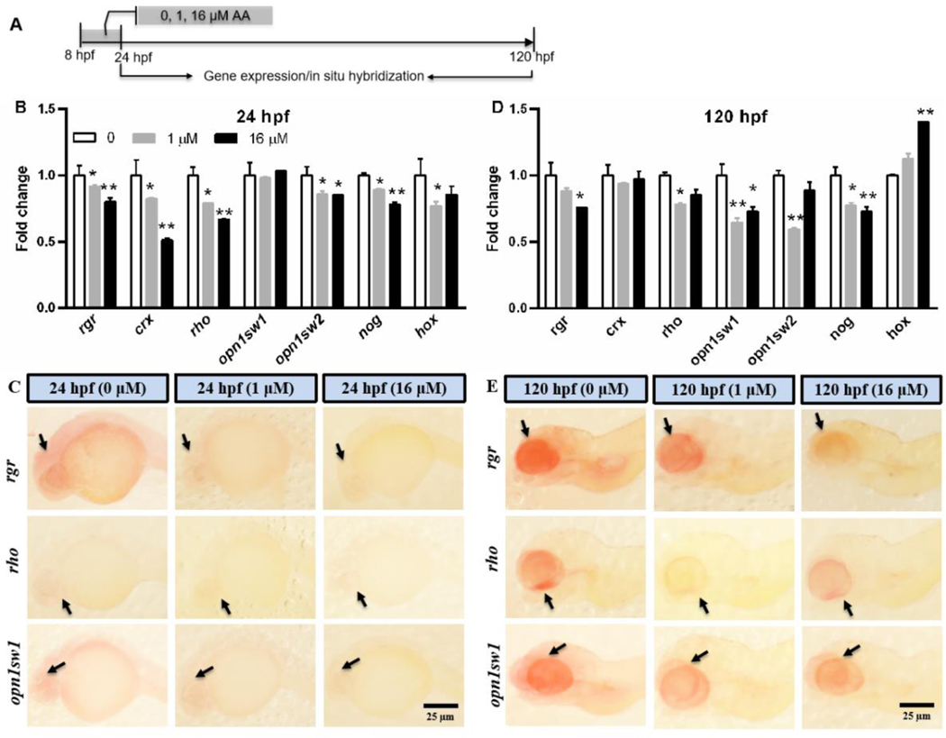
Fig. 7 Window-specific AA exposure (8–24 hpf) induced transcriptional changes in genes related to vision and motor neuron development. (A) A schematic diagram showing zebrafish embryos exposed to 0, 1, and 16 μM AA from 8–24 hpf and sampled at 24 hpf and 120 hpf for gene expression analysis. (B-E) Gene expression fold change measured by qPCR (B, D) (n = 3, each replicate consists of tissue samples pooled from 40 larvae) and spatial visualization of rgr, rho, opn1sw1 by in situ hybridization (C, E) for embryos at 24 hpf and 120 hpf. rgr: retinal G protein coupled receptor; crx: cone-rod homeobox protein; rho: rhodopsin, also known as visual purple; opn1sw1: opsin 1 (cone pigments), short-wave-sensitive 1; opn1sw2: opsin 1 (cone pigments) short-wave-sensitive 2; nog: noggin; hox: homeobox gene. Values are plotted as mean + SD. *P < 0.05 and **P < 0.001 indicate significant differences from the vehicle control (0.1 % DMSO). Arrows in (C, E) indicate gene expression position in the eye region.