
|
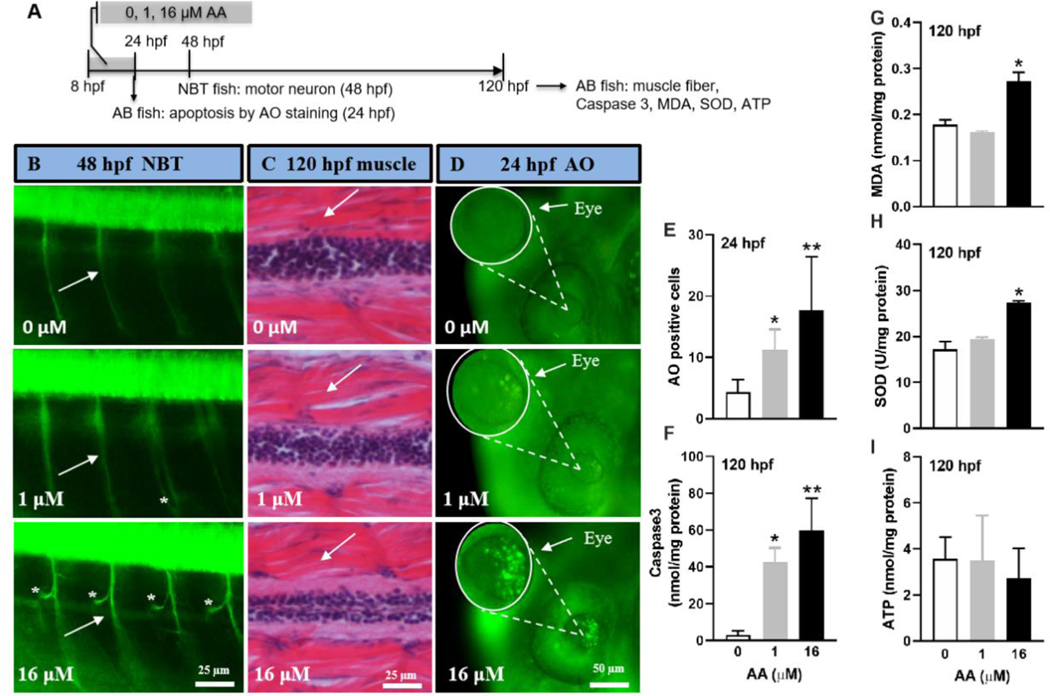
Fig. 6 Window-specific AA exposure (8–24 hpf) induced aberrant axonal growth, cell apoptosis, and oxidative stress, but had no effect on muscle fiber. (A) A schematic diagram showing zebrafish embryos exposed to 0, 1, and 16 μM AA from 8–24 hpf and sampled at various time for different endpoints. (B) Representative images showing axonal growth (white arrows) in the primary motor neuron in Tg (NBT: MAPT-GFP) fish at 48 hpf. Asterisks indicate axons with extra proliferated branches. (C) Representative histological sections of muscle fibers (white arrows) from wild type AB line fish at 120 hpf. (D) Representative images showing apoptotic cells in the eye region measured by acridine orange (AO) staining at 24 hpf. (E) Quantitative analysis of AO positive cells in (D) (n = 16–24). (F) Cell apoptosis measured by Caspase 3 in wild type AB line fish at 120 hpf. (G-I) Oxidative stress measured by MDA (G), SOD (H) and ATP (I) in wild type AB line fish at 120 hpf. For F-I, n = 3 with each replicate consists of tissue samples pooled from 20 larvae. Values are plotted as mean + SD. *P < 0.05 and **P < 0.001 indicate significant differences from the vehicle control.