
|
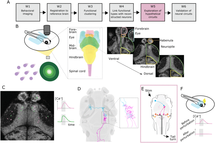
Fig. 2 Overall workflow for uncovering neural circuits – exemplified by circuits controlling movement behavior of larval zebrafish following visual stimuli. For a complete description of the steps, see Section III; note that for step W2 there is no subfigure here. A: All major working steps. B: First, a behavior imaging experiment is performed: (left) simplified view of the experimental setup and two types of stimuli: coherent dot motion stimuli (left purple) and looming stimuli (right, green); (mid) schematic drawing of relevant brain regions; (right) the activity of neural cells is recorded with Ca 2+ imaging in multiple z planes. C: The bodies of active neurons are segmented and functionally characterized based on their activity curves. D: From a large set of reconstructed neurons, those spatially closest to the active cells are determined – assuming that with some probability that they belong to the same functional type. E: Based on all the available information and pre-existing hypotheses about neural circuits, data-compatible circuits are constructed and explored. F: For the most likely hypothetical circuit, validation steps are performed, such as experimental perturbation.