
Image
|
Figure Caption
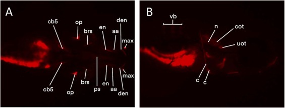
Fig. 2 Zebrafish larvae at 5 dpf with bone structures that were stained with the alizarin red live staining. Left panel (A) shows a ventral view. Right panel (B) shows a lateral view. Each structure is indicated in only one of the orientations, namely in the orientation where it was scored. The abbreviations are depicted in Table 2.
Acknowledgments
This image is the copyrighted work of the attributed author or publisher, and
ZFIN has permission only to display this image to its users.
Additional permissions should be obtained from the applicable author or publisher of the image.
Reprinted from Reproductive toxicology (Elmsford, N.Y.), 127, Hoyberghs, J., Ball, J., Trznadel, M., Beekhuijzen, M., Burbank, M., Wilhelmi, P., Muriana, A., Powles-Glover, N., Letamendia, A., Van Cruchten, S., Biological variability hampers the use of skeletal staining methods in zebrafish embryo developmental toxicity assays, 108615, Copyright (2024) with permission from Elsevier. Full text @ Reprod. Toxicol.