
|
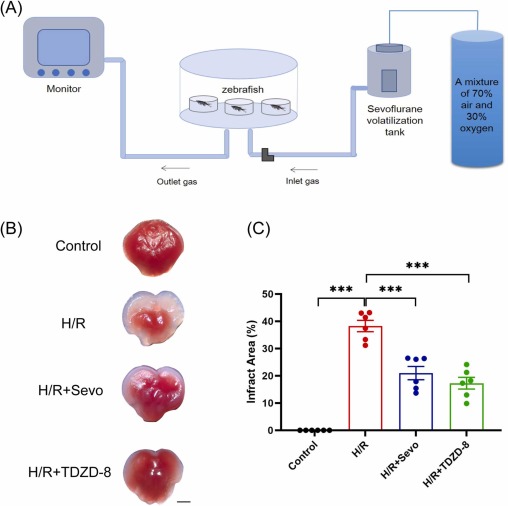
Fig. 2 Establishment of a sevoflurane postconditioning model for zebrafish and cerebral infarction detected by TTC staining. (A) The sevoflurane inhalation device for zebrafish. (B) TTC-stained brain sections of zebrafish in the control, H/R, H/R+Sevo, and H/R+TDZD-8 groups. The white regions represent the infarct area, whereas the red regions represent the non-infarct area. Scale bar = 1 mm. (C) Statistical analysis of the infarct area/total cerebral area in the four groups. Six brains in each group were analyzed. Data are shown as mean ± S.E.M. The significance level was *** p< 0.001, with analyzed by one-way ANOVA followed by Tukey’s multiple comparison test.