
|
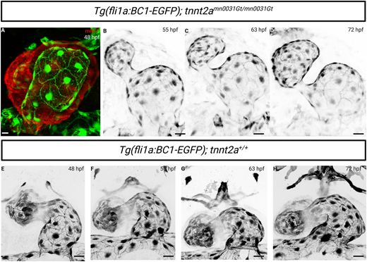
Fig. 4 Endocardial expression of the endothelial-specific β-catenin chromobody in tnnt2a mutants. (A) Confocal imaging of a 48 hpf Tg(fli1a:EGFP-BC1); tnnt2a−/− atrium; mRFP expression corresponds to the tnnt2a gene trap expression. The endothelial-specific β-catenin chromobody is localized at endocardial cell-cell junctions in tnnt2a−/−. (B-D) Confocal time-lapse images of a Tg(fli1a:EGFP-BC1); tnnt2−/− atrium at 55, 63 and 72 hpf from Movie 3. Localization of the endothelial-specific β-catenin chromobody is maintained at endocardial cell-cell junctions despite the lack of blood flow. (E-H) Confocal time-lapse images of a Tg(fli1a:EGFP-BC1); tnnt2a+/+ atrium at 48, 55, 63 and 72 hpf. Scale bars: 20 μm