
|
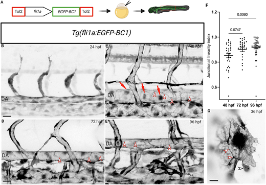
Fig. 1 The endothelial-specific β-catenin chromobody exhibits expression in endothelial nuclei and cell-cell junctions and can be used to monitor junctional maturation during zebrafish development. (A) Schematic of the fli1a:EGFP-BC1 construct. (B-E) Trunk vasculature of Tg(fli1a:EGFP-BC1) zebrafish from 24 to 96 hpf. Chromobody expression localizes to endothelial nuclei (red arrows) and at cell membranes (red arrowheads). (F) Junctional linearity index at 48 (n=32 junctions from six embryos), 72 (n=30 junctions from six larvae) and 96 (n=44 junctions from seven larvae) hpf. Data are mean±s.d.; P-value calculated using an unpaired two-tailed Student's t-test. (G) Single plane spinning disc confocal image of a single endothelial cell during CCV formation at 36 hpf. The β-catenin chromobody exhibits a dynamic localization (see Movie 1), especially at endothelial cell-cell junctions; interactions of red blood cells with endothelial cells can also be observed (red arrowheads point to endothelial cell-cell junctions; black arrowhead points to a red blood cell interacting with an endothelial cell). Scale bars: 40 μm (B-E); 10 μm (G).