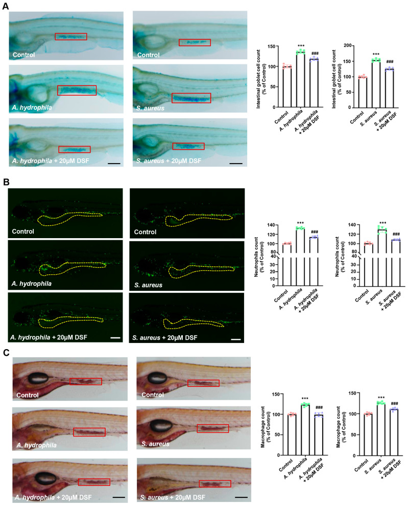
DSF restrained intestinal damage caused by the infection of A. hydrophila or S. aureus in zebrafish. (A) Imaging of Alcian blue staining and counting of goblet cells in the intestine of zebrafish (red box). (B) Live imaging of Tg(mpx:eGFP) zebrafish larva infected with A. hydrophila or S. aureus and quantitative analysis of neutrophils. (C) Imaging of macrophage accumulation (red boxes) and the quantitative analysis of macrophages in the intestine of zebrafish (red box). Data are shown as mean ± S.D. *** p < 0.001 vs. control group; ###p < 0.001 vs. A. hydrophila group or S. aureus group. Scale bar, 100 μm.
Acknowledgments
This image is the copyrighted work of the attributed author or publisher, and
ZFIN has permission only to display this image to its users.
Additional permissions should be obtained from the applicable author or publisher of the image.
Full text @ Nutrients
Your Input Welcome
Thank you for submitting comments. Your input has been emailed to ZFIN curators who may contact you if
additional information is required.
Oops. Something went wrong. Please try again later.