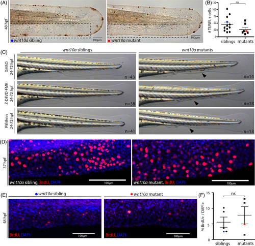
Fig. 7 MFF collapse in wnt10a mutant does not involve increased cell death or impaired cell proliferation. (A) TUNEL staining shows no major differences in the number of dying cells in the MFF of wnt10a mutant embryos at 48 hpf. (B) Quantification of the number of TUNEL-positive cells per embryo within region framed in (A), indicating no statistically significant increase in mutants compared to siblings. The blue dot and red square indicate values for embryos shown in (A). (C) Treatment from 24 to 72 hpf with the Caspase-3 inhibitor Z-DEVD-FMK (80 μM), the p53 inhibitor Pifithrin-α (0.6 μM), and DMSO (control) does not prevent the MFF collapse of wnt10a mutants at 72 hpf. (D, E) BrdU incorporation (in red) indicates no obvious differences in cell proliferation within the ventral MFF at 37 hpf, when MFF collapse commences (D), or at 48 hpf ((E), nuclei stained with DAPI in blue), when MFF is in progress (E). (F) Percentages of BrdU-positive cells in ventral MFF within the region shown in (E), indicating no statistically significant decrease in mutants compared to siblings. The blue dot and red square indicate values for embryos shown in (E). MFF, median fin fold.
Image
Figure Caption
Acknowledgments
This image is the copyrighted work of the attributed author or publisher, and
ZFIN has permission only to display this image to its users.
Additional permissions should be obtained from the applicable author or publisher of the image.
Full text @ Dev. Dyn.

