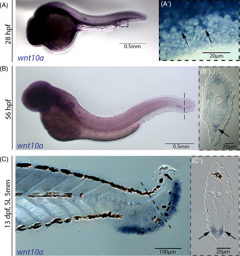
Fig. 5 wnt10a is expressed in fin mesenchymal cells. wnt10a in situ hybridization of wild-type embryos. (A) At 28 hpf, wnt10a is expressed in the fin mesenchymal cells, as the cells start to migrate into the dermal space of the MFF (arrows in A', showing a magnified view of region framed in (A)). (B) At 56 hpf, wnt10a is expressed in fin mesenchymal cells throughout the entire dermal space of the MFF (arrow in B′, showing a transverse section at the position indicated by dashed line in (B)). (C) At the onset of metamorphosis at 13 dpf (SL 5 mm), wnt10a is strongly expressed in the dermal space of the caudal fin where lepidotrichia start to form (arrows in (C′), showing a transverse section at the position indicated by dashed line in (C)). MFF, median fin fold; SL, standard length.
Image
Figure Caption
Acknowledgments
This image is the copyrighted work of the attributed author or publisher, and
ZFIN has permission only to display this image to its users.
Additional permissions should be obtained from the applicable author or publisher of the image.
Full text @ Dev. Dyn.

