
|
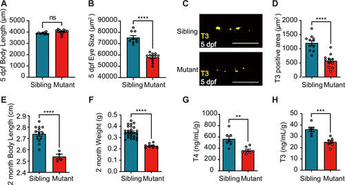
Fig. 2 031003 mutant presented thyroid dysgenesis phenotypes. A: the body length of the 031003 sibling vs. mutant at 5 days postfertilization (dpf) (ns: no significant difference). B: eye size of the 031003 sibling vs. mutant at 5 dpf. C: confocal examination of mature thyroid follicles marked by triiodothyronine (T3) immunofluorescence at 5 dpf. Scale bar: 100 μm. D: the areas of mature thyroid follicles per embryo were quantified by Imaris software. Whole body length (E) and weight (F) of 2-mo-old adult 031003 zebrafish. G and H: thyroid hormone levels in 2-mo-old adult zebrafish by ELISA. The sera of three individuals were pooled in a tube, and each group was analyzed in triplicate. **P < 0.01, ***P < 0.001, ****P < 0.0001. T4, thyroxine.