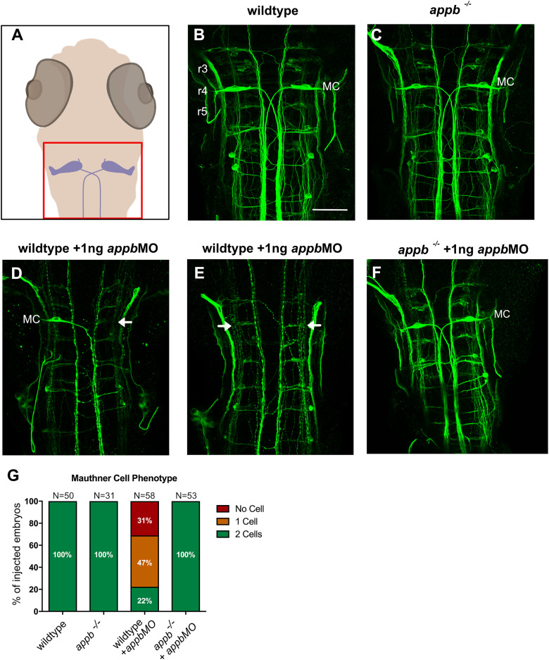
appbMO-mediated loss of Mauthner cells in wild type but not appb−/− zebrafish. A, Schematic image of the hindbrain region shown in B–F. B, C, Dorsal view of hindbrain of embryos (anterior to the top) stained with RMO44 antibody at 48 hpf, displaying the large Mauthner cells (MC) in wild type (B) and appb−/− (C). D, E, Wild-type larvae injected with 1 ng splice-blocking appbMO showed two (22%), one (47%, D), or no MC (31%, E). F, two MC were observed in all appb−/− injected with 1 ng splice-blocking appbMO. G, Quantification of the MC number at 48 hpf. G, n = 3 biologically independent samples. “N” indicates number of brains. r3–r5, rhombomeres 3–5. Scale bar, 50 μm. MC, Mauthner cells. Arrows indicate missing Mauthner cells.
This image is the copyrighted work of the attributed author or publisher, and
ZFIN has permission only to display this image to its users.
Additional permissions should be obtained from the applicable author or publisher of the image.
Full text @ eNeuro
Your Input Welcome
Thank you for submitting comments. Your input has been emailed to ZFIN curators who may contact you if
additional information is required.
Oops. Something went wrong. Please try again later.