
|
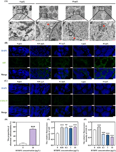
Fig. 5 Potential effects on mitochondria in zebrafish testes upon BTBPE exposure. (A) Mitochondrial ultrastructure (a’: 0 μg/L, while b’–d’: 10 μg/L. a’–d’ are magnified views of a–d, respectively. Scale bar = 500 nm). Mitochondrial immunofluorescence of (B) apoptosis-inducing factor 1 (AIF) and (C) cytochrome c oxidase subunit 4 isoform 1 (COX IV). Scale bar = 50 μm. (D) The proportion of abnormal mitochondria increases upon BTBPE treatments. The relative fluorescence intensity of (E) AIF and (F) COX IV. Data represented as the mean ± SEM (n = 3 individuals). **p < 0.01 and ***p < 0.001 denote statistically significant differences between BTBPE treatment and the control.