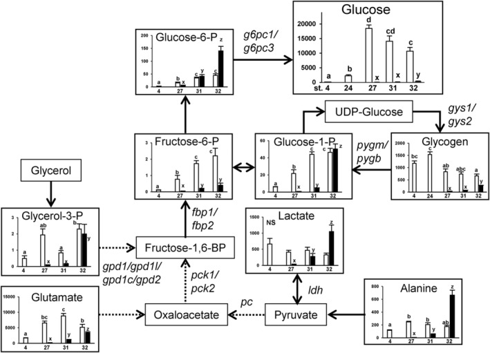
IMAGE

FIGURE 1

ID
ZDB-IMAGE-240601-9
Source
Figures for Shimizu et al., 2024
Image
Figure Caption

FIGURE 1

The metabolic pathway map showing changes in each metabolite levels per individual yolk sac (open column) or embryo (filled column) during development. The horizontal axes represent developmental stages {stages 4, 24 (glucose and glycogen only), 27, 31, 32} and the vertical axes represent nmol/sample. Data are presented as mean ± standard error (N = 6). Different letters indicate significant differences (p < 0.05) between groups. Tests for significant differences were performed by one‐way ANOVA and Tukey's post‐hoc test separately for yolk sac or embryo samples after log transformation. Fructose‐1,6BP, fructose 1,6‐bisphosphate; ‐P, ‐phosphate. Genes responsible for respective pathways were shown beside the arrows. For the complete map with all metabolites measured in this study, please refer to Figure S2.

Acknowledgments
This image is the copyrighted work of the attributed author or publisher, and ZFIN has permission only to display this image to its users. Additional permissions should be obtained from the applicable author or publisher of the image. Full text @ Physiol. Rep.