IMAGE

FIGURE 4

ID
ZDB-IMAGE-240601-11
Source
Figures for Shimizu et al., 2024
Image
Figure Caption

FIGURE 4

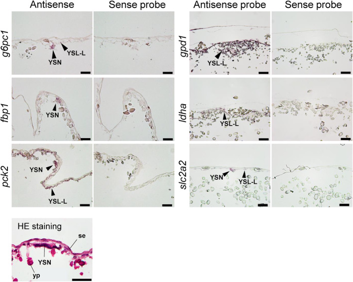
In situ hybridization analysis for g6pc1, fbp1, pck2, gpd1, ldha, and slc2a2 at yolk sac membrane (YSM) at stage 27. For all transcripts, sense probes (right) were used as negative controls for antisense probes (left), which showed true signals. Positive signals for transcripts are indicated by arrowheads. Hematoxylin and eosin (HE) staining shows general morphology of the YSM in stage 27. se, squamous epithelium; yp, yolk platelet; YSN, yolk syncytial nuclei. Scale bars = 50 μm.

Acknowledgments
This image is the copyrighted work of the attributed author or publisher, and ZFIN has permission only to display this image to its users. Additional permissions should be obtained from the applicable author or publisher of the image. Full text @ Physiol. Rep.